






Brief Description
This SOP describes the procedure for determining particle size using microscopy in the Quality Control laboratory. It is intended for microscopic characterization of particles generally greater than 1 µm, using a compound microscope fitted with a calibrated ocular measuring scale. For analysis, approximately 10–100 mg of sample is dispersed in 10 mL of a suitable non-dissolving suspending medium, such as paraffin oil or glycerin. Two to three drops are placed on a clean glass slide, covered with a cover slip, and examined under 10X, 40X, or 100X magnification. Particle dimensions are measured using the ocular scale, and observations are made in at least five different microscopic fields. The SOP also covers particle-size parameters such as Feret’s diameter, Martin’s diameter, projected-area diameter, length and width, together with particle-shape and surface characterization. Particle size in microns is calculated as number of scale divisions × lens factor, using factors of 10, 2.5 and 1 µm for 10X, 40X and 100X, respectively. Results are recorded on the specified test data sheet and reported as a percentage.
Skip to PDF content1. Flow Diagram:
The flow diagram presents the step-by-step procedure for particle size analysis using a calibrated compound microscope in the Quality Control laboratory. The process begins by taking 10–100 mg of sample and preparing a homogeneous suspension in 10 mL of a suitable non-dissolving medium, such as paraffin oil or glycerin. Two to three drops of the suspension are placed on a clean glass slide and covered with a cover slip.
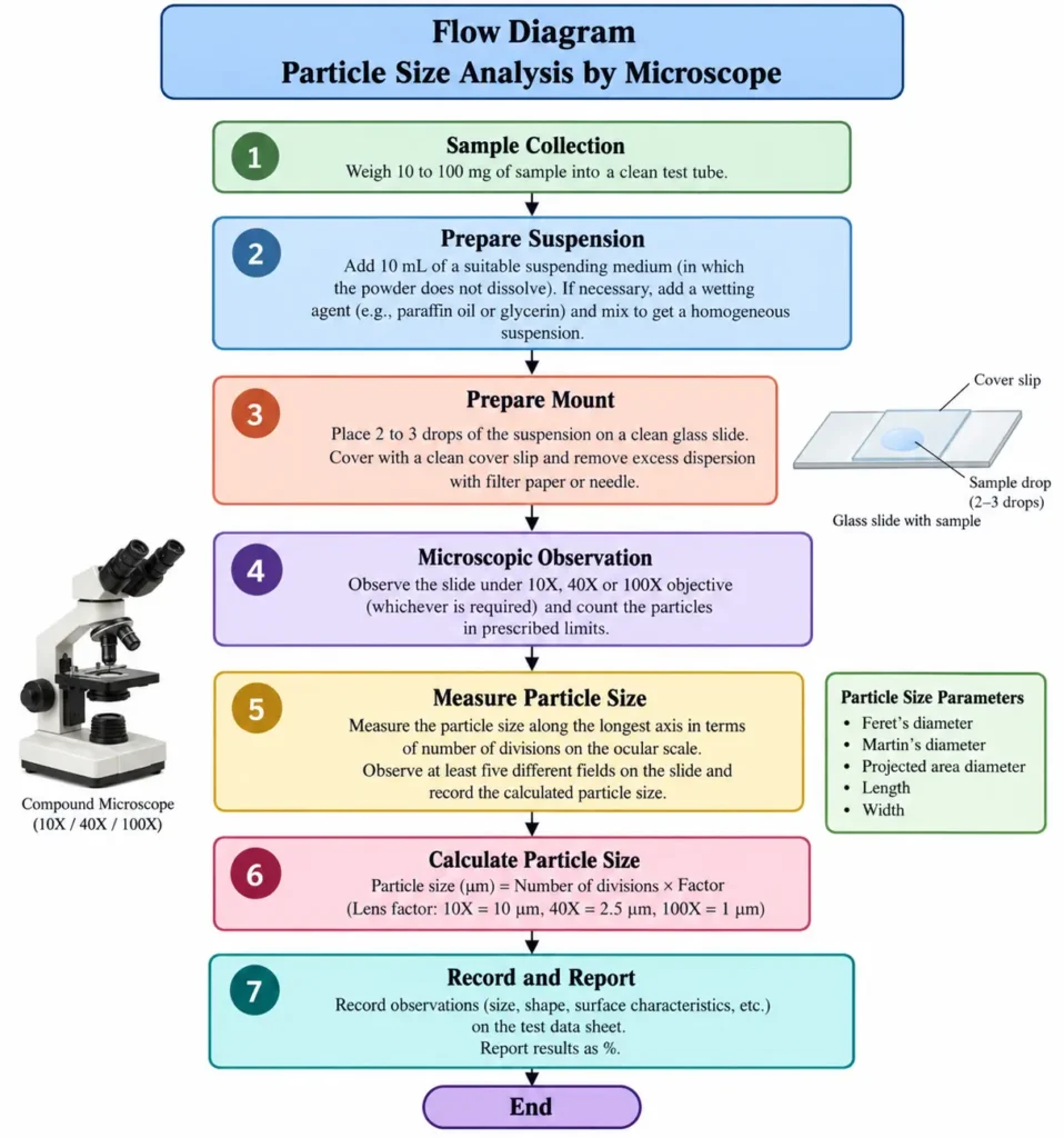
The prepared slide is then observed under 10X, 40X, or 100X magnification, depending on the requirement. Particle size is measured along the longest axis using the ocular scale, with observations taken from at least five different microscopic fields.The particle size is calculated using number of scale divisions × lens factor, with factors of 10, 2.5, and 1 µm for 10X, 40X, and 100X, respectively. The observations, particle characteristics, calculations, and final percentage results are documented in the test data sheet.
2. Brainstorming Diagram for SOP Failure:
The brainstorming diagram identifies the major causes that may lead to ineffective implementation of the SOP for Particle Size Analysis by Microscope in a pharmaceutical manufacturing/QC environment. The potential causes are grouped under Man, Machine, Method, Material, Measurement, Environment, Management, Miscellaneous, Manpower Motivation, and Regulatory/Quality factors.
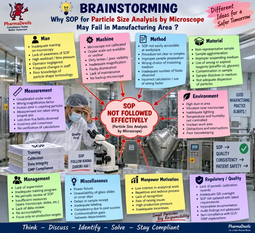
Key risks include inadequate analyst training, lack of SOP awareness, uncalibrated microscope or ocular scale, dirty lenses, incorrect sample preparation, unsuitable suspending medium, poor particle dispersion, wrong magnification factor, insufficient microscopic fields, calculation errors, poor supervision, inadequate documentation, and weak QA oversight. These factors can directly affect the reliability and reproducibility of particle-size results. The SOP specifically requires a calibrated microscope, representative sample preparation, suitable mounting medium, observation under appropriate magnification, measurement from at least five different fields, and correct use of lens factors for calculation. The diagram therefore emphasizes that proper training, calibration, supervision, documentation, sample handling, and GMP/GLP compliance are essential to prevent SOP failure and ensure accurate microscopic particle-size analysis.
3. 5-Why Analysis for SOP Failure:
The 5-Why analysis identifies the underlying reasons why the SOP for Particle Size Analysis by Microscope may not be followed effectively in the manufacturing/QC area. The analysis begins with the immediate failure that the analyst did not perform the microscopy procedure according to the SOP requirements for sample preparation, microscopic observation, particle measurement, and calculation.
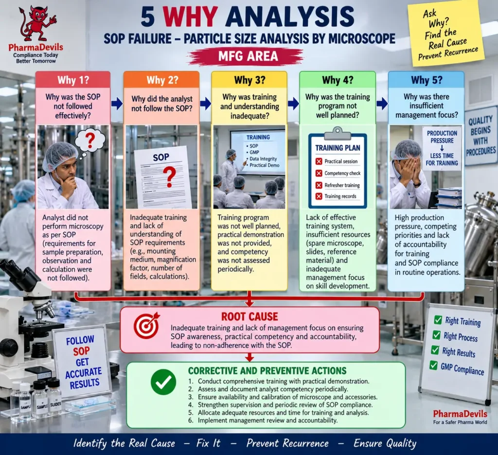
The successive “Why” questions indicate that the failure may arise from inadequate training, poor understanding of SOP requirements, lack of practical demonstration, insufficient competency assessment, inadequate training planning, limited resources, production pressure, and weak management focus on SOP compliance. This is significant because the SOP requires correct sample dispersion, use of suitable mounting medium, observation under the required magnification, measurement from at least five fields, and proper particle-size calculation. The identified root cause is therefore insufficient training, competency control, supervision, and management attention, which can lead to inconsistent execution and unreliable particle-size results. Appropriate CAPA should include practical training, periodic competency assessment, calibrated equipment, adequate resources, strengthened supervision, and routine review of SOP compliance.
4. Fishbone Analysis for SOP Failure:
The fishbone diagram identifies the major potential causes for failure to effectively follow the SOP for Particle Size Analysis by Microscope in the manufacturing/QC area. The causes are grouped into Man, Machine, Method, Material, Environment, Measurement, and Management categories.
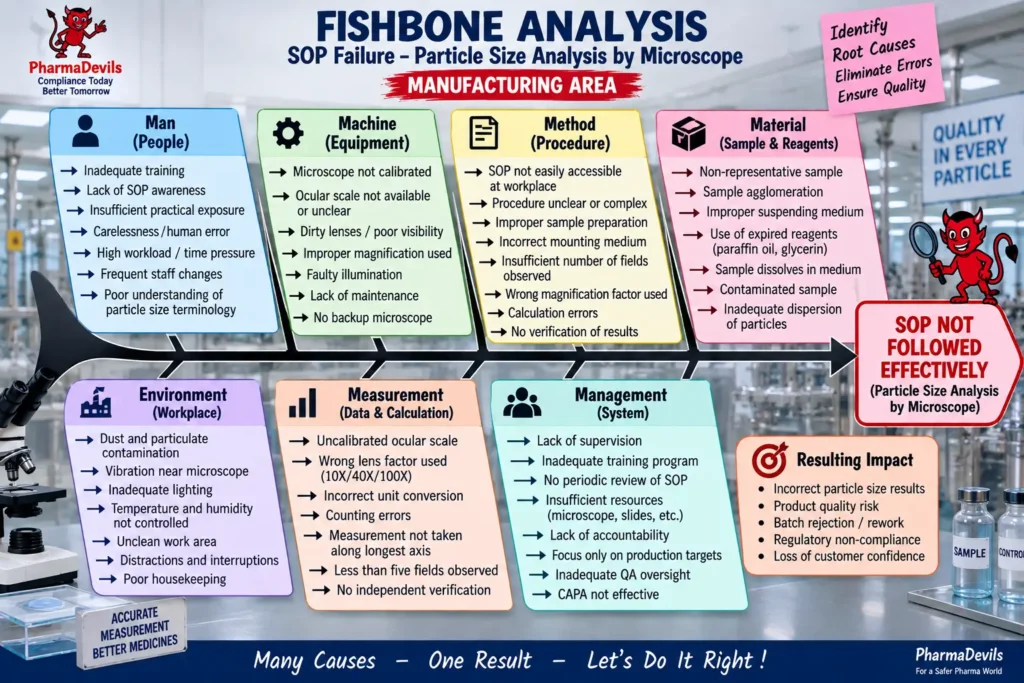
Key issues include inadequate analyst training, lack of SOP awareness, uncalibrated microscope or ocular scale, dirty lenses, incorrect magnification, improper sample preparation, unsuitable mounting or suspending medium, non-representative samples, inadequate particle dispersion, poor environmental conditions, wrong lens factor, counting or calculation errors, insufficient supervision, inadequate resources, and weak QA oversight. These factors can interfere with the SOP requirements for representative sample preparation, microscopic observation at the required magnification, measurement from at least five fields, and correct particle-size calculation. The analysis shows that SOP failure is usually multifactorial rather than caused by a single error. Effective control therefore requires proper training, calibrated equipment, standardized sample preparation, accurate calculations, adequate supervision, and periodic review of compliance to ensure reliable particle-size results.
5. Fault Tree Analysis for SOP Failure:
The Fault Tree Analysis illustrates how different failures can combine to cause ineffective implementation of the SOP for Particle Size Analysis by Microscope in the manufacturing/QC area. The top event is SOP not followed effectively, resulting in incorrect or inconsistent particle-size results. The diagram groups the possible causes into analyst-related, equipment-related, method-related, material-related, environment-related, measurement/calculation-related, and management-related failures. Examples include inadequate training, lack of SOP awareness, uncalibrated microscope, unclear ocular scale, dirty lenses, improper sample preparation, incorrect mounting medium, insufficient fields observed, non-representative sample, sample agglomeration, wrong lens factor, counting errors, poor supervision, and inadequate QA oversight.
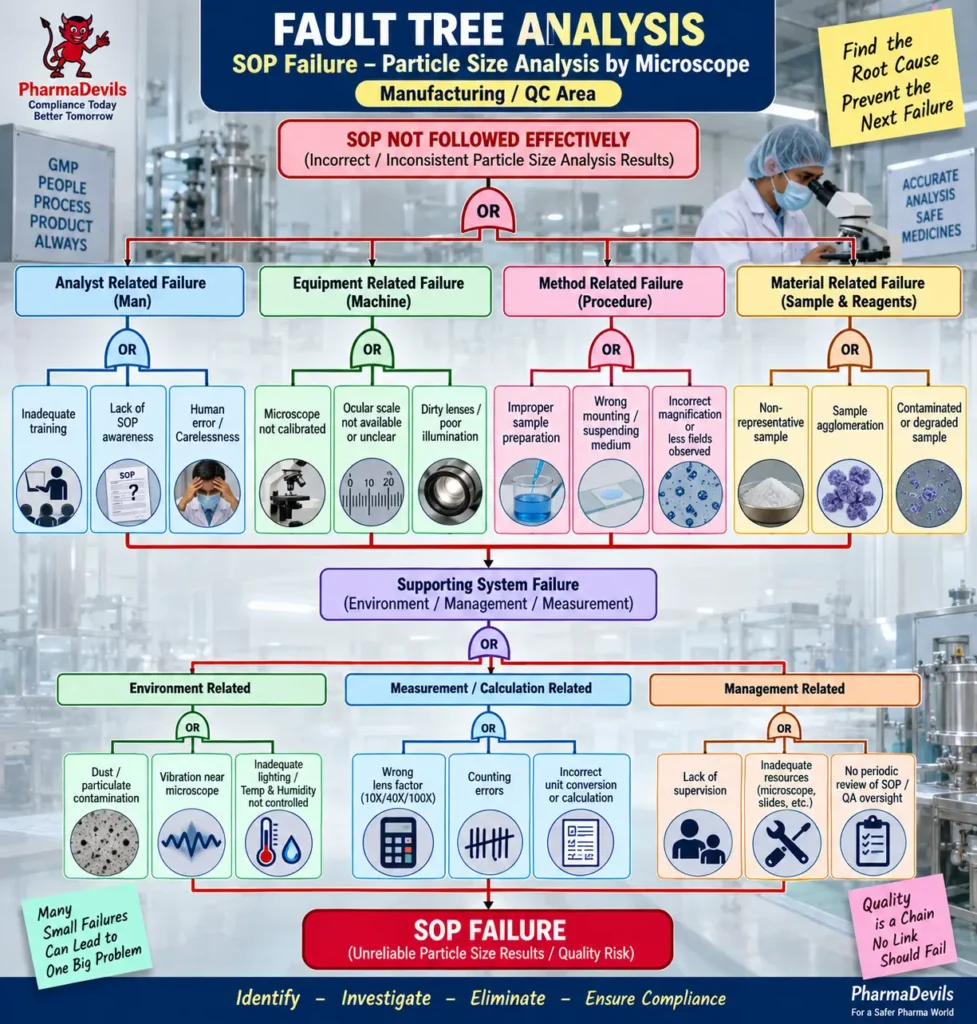
These causes are relevant because the SOP requires suitable sample dispersion, proper slide preparation, observation under 10X, 40X, or 100X, measurement from at least five different fields, and correct use of the specified lens factors during calculation. The analysis shows that several small control failures may collectively lead to unreliable particle-size results, product-quality risk, rework or rejection, and GMP non-compliance. The purpose of the fault tree is to systematically identify these failure pathways and support effective CAPA and recurrence prevention.
Questions & Answers – Particle Size Analysis by Microscope
- Q: What is the objective of this SOP?
A: To describe the procedure for determining particle size using microscopy in the Quality Control laboratory. - Q: Who is responsible for performing the particle size analysis?
A: The Chemist-QC is responsible for executing the procedure, while the Section Head, Head-QC, and Lab QA are responsible for ensuring and monitoring compliance with the SOP. - Q: For what particle size range is microscopy considered suitable in this SOP?
A: The SOP states that microscopic particle-size analysis is suitable for particles greater than approximately 1 µm. - Q: What equipment is required for the analysis?
A: A compound microscope with a calibrated ocular measuring scale, along with clean glass slides and cover slips. - Q: What quantity of sample is used for particle size determination?
A: Approximately 10–100 mg of sample is transferred into a test tube. - Q: How much suspending medium is added to the sample?
A: 10 mL of a suitable suspending medium in which the powder does not dissolve is added. - Q: Which mounting or suspending media are mentioned in the SOP?
A: The SOP mentions liquid paraffin and glycerin as suitable media, depending on the properties and solubility of the sample. - Q: How is the microscopic slide prepared?
A: About 2–3 drops of the prepared suspension are placed on a clean glass slide, covered with a clean cover slip, and excess dispersion is removed using filter paper or a needle. - Q: Which microscope objectives may be used?
A: The sample may be observed under 10X, 40X, or 100X, depending on the requirement. - Q: How should particle size be measured?
A: Particle size is measured along the longest axis in terms of the number of divisions on the ocular scale. - Q: How many microscopic fields should be observed?
A: Particle size should be observed from at least five different fields on the slide. - Q: What particle-size parameters are described in the SOP?
A: The SOP describes Feret’s diameter, Martin’s diameter, projected-area diameter, length, and width. - Q: What particle shapes are mentioned?
A: Examples include acicular, columnar, flake, plate, lath, and equant particles. - Q: How is particle size in microns calculated?
A:
Particle Size (µm) = Total number of divisions × Factor.
The factors stated are 10 for 10X, 2.5 for 40X, and 1 for 100X. - Q: How are the results reported?
A: The SOP specifies that the result should be reported as a percentage and recorded on the test data sheet. - Q: What is the calibration requirement for the microscope?
A: The microscope is to be calibrated according to the referenced instrument calibration SOP, /QC/INS/69-00. - Q: Who provides training for this SOP?
A: The Manager-QC is the trainer, the Chemist-QC is the trainee, and the training period specified is one hour. - Q: Why is proper dispersion of particles important?
A: The particles should be adequately dispersed so individual particles can be distinguished and the observed particles remain representative of the material’s size distribution.
Reference Guidelines:
- Indian Pharmacopoeia (IP) – General Chapter 2.5.8: Optical Microscopy: Particle Size by Microscopy. The Indian Pharmacopoeia Commission identifies this as the harmonized chapter for optical microscopy and notes its implementation from 1 December 2022. (Indian Pharmacopoeia Commission)
- USP General Chapter <776> – Optical Microscopy. This chapter addresses microscopic characterization and measurement of particles and is part of the Pharmacopoeial Discussion Group harmonization work. (USP)
- European Pharmacopoeia – Chapter 2.9.37, Optical Microscopy. The current Ph. Eur. index includes Chapter 2.9.37 Optical Microscopy, corresponding to the harmonized optical microscopy procedure. (EDQM)
- WHO TRS 1052, Annex 4 – Good Practices for Pharmaceutical Quality Control Laboratories (2024). This guideline covers laboratory quality systems, personnel, equipment, documentation, testing procedures, records, and controls needed to obtain accurate and reliable QC results. (World Health Organization)
- ISO/IEC 17025:2017 – General Requirements for the Competence of Testing and Calibration Laboratories. WHO’s current pharmaceutical QC laboratory guideline specifically states consistency with ISO/IEC 17025:2017, making it relevant for microscope calibration, analyst competency, method control, and reliable test results. (World Health Organization)