









Step-by-Step Procedure
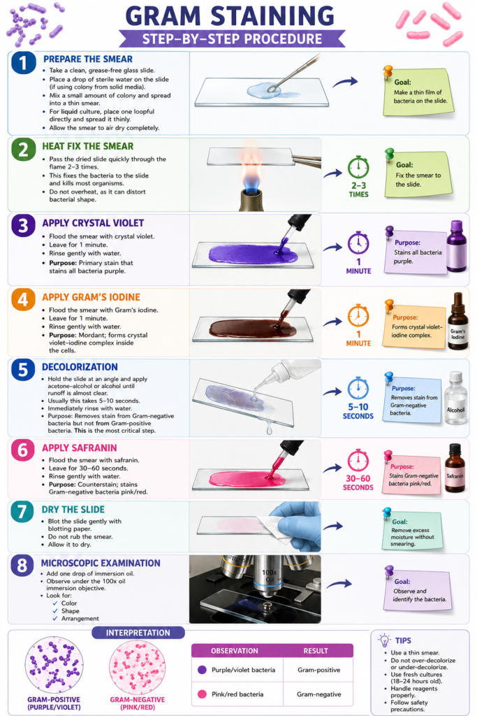
1. Prepare the Smear
Take a clean, grease-free glass slide.
Place a small drop of sterile water on the slide if using a colony from solid media.
Mix a small amount of bacterial colony into the drop and spread it into a thin smear.
For liquid culture, place one loopful directly on the slide and spread it thinly.
Allow the smear to air dry completely.
2. Heat Fix the Smear
Pass the dried slide quickly through the flame 2–3 times.
This fixes the bacteria to the slide and kills most organisms.
Do not overheat, as it can distort bacterial shape.
3. Apply Crystal Violet
Flood the smear with crystal violet.
Leave for 1 minute.
Rinse gently with water.
Purpose: Primary stain that stains all bacteria purple.
4. Apply Gram’s Iodine
Flood the smear with Gram’s iodine.
Leave for 1 minute.
Rinse gently with water.
Purpose: Mordant; forms crystal violet–iodine complex inside the cells.
5. Decolorization
Hold the slide at an angle and apply acetone-alcohol or alcohol until runoff is almost clear.
Usually this takes 5–10 seconds.
Immediately rinse with water.
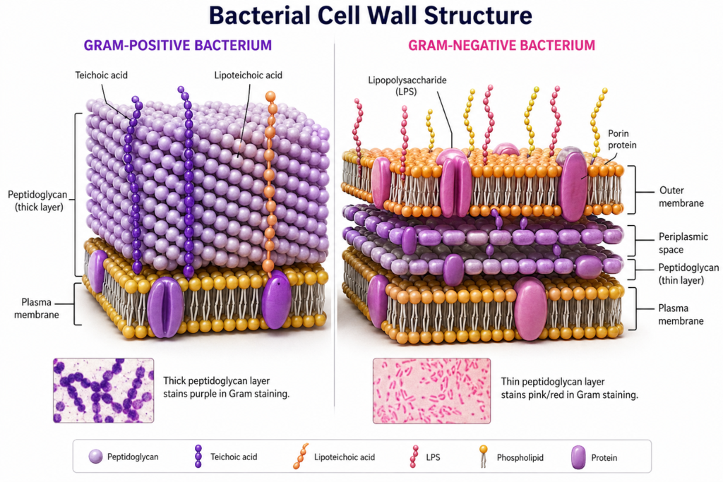
Purpose: Removes stain from Gram-negative bacteria but not from Gram-positive bacteria.
This is the most critical step.
6. Apply Safranin
Flood the smear with safranin.
Leave for 30–60 seconds.
Rinse gently with water.
Purpose: Counterstain; stains Gram-negative bacteria pink/red.
7. Dry the Slide
Blot the slide gently with blotting paper.
Do not rub the smear.
Allow it to dry.
8. Microscopic Examination
Add one drop of immersion oil.
Observe under the 100x oil immersion objective.
Look for:
- Color
- Shape
- Arrangement
Interpretation
| Observation | Result |
|---|---|
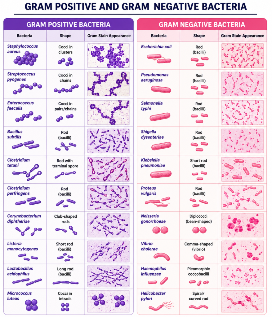
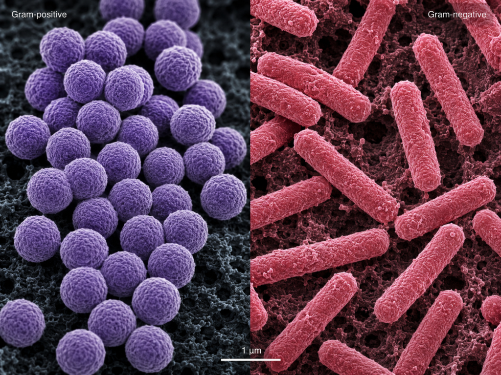
| Purple/violet bacteria | Gram-positive |
| Pink/red bacteria | Gram-negative |